Two studies published in 2026 reported significant reductions in pain and disability after DRX9000 non-surgical spinal decompression. For patients living with chronic low-back pain, the clearest finding was an improvement in symptoms and everyday function.[1][2]
In the first study, average pain scores decreased by approximately 61%, from 5.6 to approximately 2.2 out of 10. The second reported an average improvement of 3.23 points on the same scale. Both also found improved scores on a questionnaire measuring how back pain affects daily activities.[1][2]
The clinical findings are encouraging: substantial pain relief and better daily function after non-surgical spinal decompression. The papers also add detailed MRI assessments of the spine before and after care.
The researchers also examined MRI scans. One study found an increase in spinal canal dimensions; the other explored how the composition of supporting muscles related to changes in function. Together, the papers provide both patient-reported outcomes and detailed measurements of the lower spine.
For a larger study focused on pain and daily activities, see our explanation of the 267-record Military Medicine study.
What is non-surgical spinal decompression?
Non-surgical spinal decompression uses computer-controlled traction to apply and release a measured pulling force through the spine. The treatment is designed to reduce mechanical loading on the spinal discs. Both studies used the DRX9000 system, with treatment settings adjusted for each patient.[1][2]
The research focused on the lumbar spine, or lower back. It examined existing treatment records and MRI scans from patients treated at USF Health.
01 / How the research was done
Using MRI to measure changes in the spine
MRI provides detailed images of spinal discs, nerves and soft tissues. In the first study, researchers used automated software to identify the boundaries of spinal structures and measure their size. This technique, called segmentation, allowed them to compare the same anatomical features before and after treatment.[1]
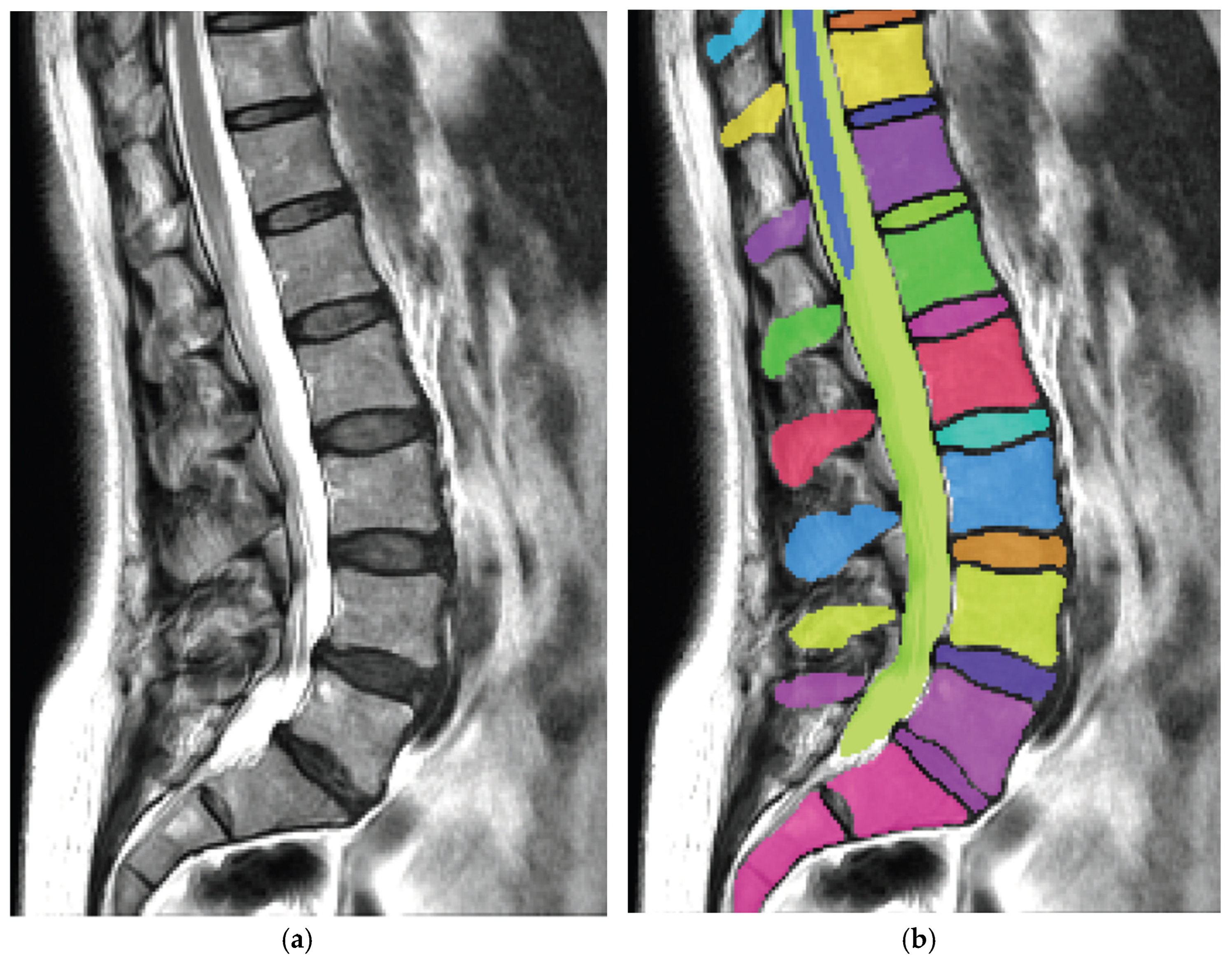
The study included 36 people with chronic low-back pain. A typical course consisted of 20 treatment sessions lasting 28 minutes each. The median time between the initial and follow-up MRI scans was approximately five months.[1]
02 / The structural study
Less pain and improved daily function
Average pain scores fell by 3.4 points on a 0-to-10 scale, from 5.6 before treatment to approximately 2.2 afterward, a reduction of about 61% in the group’s average score. Among the 20 participants with follow-up disability scores, the average improvement was 10.8 points on the study’s 0-to-50 Oswestry Disability Index.[1]
The Oswestry questionnaire measures how back pain affects activities such as walking, sitting, sleeping and personal care. These scores help describe the practical effect of symptoms on daily life, alongside their intensity.
Small increases in spinal canal dimensions
The spinal canal is the passage within the spine that contains neural structures. At the treated level, its average cross-sectional area increased from 170.9 to 175.8 square millimeters, a change of 2.9%. Its average side-to-side diameter increased from 16.0 to 16.3 millimeters.[1]
Spinal canal area · 36 participants
A modest change in measured space
Increase in average spinal canal area
at the treated level.
The MRI findings add an objective structural measurement to the clinical results. A small increase in spinal canal area was documented alongside substantially lower pain scores and improved daily function.
What happened to the discs?
Disc thickness did not significantly increase across the group, and average disc volume decreased by approximately 4%. In participants treated at L5, measured disc thickness decreased. The area of the openings where nerves exit the spine did not significantly change.[1]
The authors proposed that changes in fluid distribution or disc shape might help explain these findings. Those mechanisms were not directly measured. The results do not demonstrate disc regeneration, but they do show that symptom improvement can occur without an increase in disc height.[1]
An earlier CT study reported increased disc height alongside less pain. Our companion article explains that result and how it differs from the newer MRI findings.
For an explanation of common terms used in MRI reports, see our guide to bulging and herniated discs.
03 / The muscle study
Changes in the muscles that support the lower back
The second study examined 21 people and used MRI analysis to measure muscle and fat around the lumbar spine. Researchers were interested in whether changes in these tissues were associated with improvements in pain and function.[2]
Average pain scores improved by 3.23 points. Disability scores improved by 10.83 points among the 12 participants with follow-up measurements. There were no statistically significant changes in muscle measurements across the full group.[2]
In a subgroup of eight women with follow-up disability scores, greater improvement in function was associated with reductions in fat area in three supporting muscles. This preliminary finding warrants further study. The sample was too small to establish differences in treatment response between women and men.[2]
04 / Putting the findings together
What do these findings mean for patients?
The main clinical result is straightforward: patients reported substantially less pain after treatment, and those with follow-up disability questionnaires reported better daily function. The MRI analysis adds information about the anatomical changes observed during that period.
The studies also highlight why treatment progress is assessed in several ways. Symptoms, activity levels, examination findings and imaging provide different information. A change in one MRI measurement does not necessarily reflect the extent of a person’s improvement.
Assessing progress
What improvement can look like
- Pain: Symptoms become less intense or interfere less often with daily life.
- Function: Walking, sitting, sleeping or other important activities become easier.
- Clinical findings: Follow-up examination helps assess progress and guide the next steps in care.
Follow-up MRI may be appropriate when it would help answer a specific clinical question. These studies do not establish a need for routine repeat scans during treatment.
The bottom line
Both studies documented substantial pain relief and improved daily function after DRX9000 treatment. The structural study also measured an increase in spinal canal dimensions. Together, the findings strengthen the clinical evidence for non-surgical spinal decompression and give patients concrete results to consider when choosing care for chronic low-back pain.
Our guide to who may be a candidate for spinal decompression explains the considerations involved in an individual evaluation.
Research references
Sources & study notes
- Connolly JA, Molinier N, Weber KA II, Cohen-Adad J, Schilaty ND. Structural Remodeling of the Lumbar Spine Following Non-Surgical Spinal Decompression: An Automated MRI Study. Journal of Functional Morphology and Kinesiology. 2026;11(4):390. Published September 29, 2026. Full text reviewed. Read the full structural MRI study.
- Connolly JA, Weber KA II, Wesselink EO, Baez A, Sun A, Schilaty ND. Sex-specific differences in paraspinal muscle composition and clinical outcomes after non-surgical spinal decompression. BMC Musculoskeletal Disorders. Published October 2, 2026. Article-in-press PDF and supplement reviewed; the final edited version may change. Read the back-muscle MRI study and supplements.
Funding: Both papers disclose support involving Excite Medical, the DRX9000 manufacturer, alongside the Florida High-Tech Corridor and the Florida Department of State Center for Neuromusculoskeletal Research. The authors report freedom to publish positive or negative results.
Study design: These small, retrospective studies had no comparison groups. MRI timing varied, some follow-up disability scores were missing, and the muscle study did not track physical activity. They do not establish superiority over other care or how long improvements last. The papers share a clinical site and research program. Patient overlap is not clarified, so the sample sizes should not be combined as independent groups. Both studies made multiple statistical comparisons without an adjustment for multiple testing; exploratory associations require confirmation.
Scope: These are lumbar studies, not evidence about cervical decompression or a comparison with surgery. Other treatments received during care are not sufficiently described to isolate their contribution. The structural paper’s abstract and results differ slightly in mean age and disc volume; this article avoids the age discrepancy and uses the results section for the imaging discussion.
Editorial disclosure: Madalian Wellness offers DRX9000 treatment. This article is prepared under the practice’s editorial responsibility and reviewed by Dr. Henry Madalian, not written by the study authors.